

Dural arteriovenous fistula (dAVF) is a relatively rare condition which usually arises after clotting in one of the large veins (dural sinuses) around the brain or spinal cord. They can also form following head injury, infection, brain surgery and in conjunction with brain or meningeal tumours. When the body’s healing mechanisms attempt to dissolve the clot and recanalise the veins, abnormal connections develop between the arteries supplying the dura (the tough, fibrous lining surrounding the brain which houses the dural sinuses) and the dural sinuses themselves. Occasionally, a dAVF develops in the large vein behind the eyes, the cavernous sinus. In such cases, the lesion is referred to as a carotid-cavernous fistula (CCF).
Cranial dural arteriovenous fistula account for around 10 – 15% of all intracranial vascular malformations. The majority of the patients are asymptomatic while some patients may experience pulsatile tinnitus (a “whooshing” noise heard in the ears in time with the pulse), headaches and visual disturbances. Brain haemorrhage, seizures, weakness, progressive confusion and dementia may also result.
Spinal dAVF can present with lower limb weakness, progressive paralysis, bladder/bowel dysfunction and sensory disturbance.
In the case of CCF, the most common symptoms are protrusion of one or both eyes, eye redness, eye pain, deterioration of vision, and occasionally pulsatile tinnitus.
Diagnosis of cranial dAVF and CCF can be difficult. Occasionally, under the keen eye of an expert neuroradiologist they can be detected on CT or MRI. However, most cases require catheter angiography (DSA) performed by an experienced neurointerventionist for definitive diagnosis.
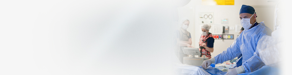
The mainstay of treatment for dAVF and CCF in the 21st century is interventional neuroradiology with embolisation (targeted blocking of abnormal vessels under image guidance). These procedures are performed under general anaesthesia. In the case of dAVF, the treatment is usually performed through a small (3-4mm) incision in the groin or wrist. In the case of CCF, treatment can be conducted through and incision in the groin or wrist, or occasionally, by inserting a small needle under the eye to puncture the abnormal veins.
We are specialists in management of dAVF and CCF and look after all aspects of dAVF care:
- Performing diagnostic catheter angiography
- Medical management and surveillance
- Neurointerventional treatments including transvenous embolisation, transarterial embolisation, percutaneous embolisation.
Dr Wenderoth has a special interest in dural arteriovenous fistula, and has pioneered and published on several techniques which have revolutionised the way dAVF is treated and led to much more robust and successful cure rates.